


Differential Tissue Harmonic Imaging of an Ovarian Dermoid Cyst
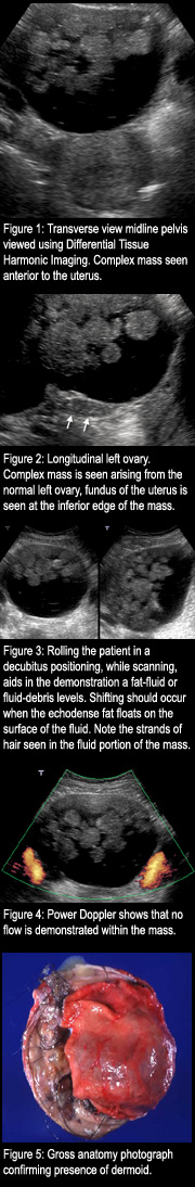
SummaryA dermoid, or mature teratoma, is a benign type of ovarian tumor. Dermoids are common, constituting about one-third of all benign ovarian tumors and often found in young women. Dermoids are bilateral in approximately 10% to 20% of cases. HistoryThis 22-year-old female, presented with a large pelvic mass on physical examination, with lack of pelvic pain. FindingsEndovaginal and trans-abdominal pelvis sonography was performed using the Aplio ultrasound system. The uterus was mildly prominent in size. Its increased size was primarily in length because of being stretched by a large midline pelvic complex cystic mass. The right ovary was normal sonographically. A large complex mass was found arising from the left ovary, that did not seem to grossly distort the left ovary but appeared to rise from the surface of the left ovary. The complex cystic mass contained globules of fat floating on fluid with stringy echoes compatible with hair, rather typical for a dermoid. Measurements of the mass could only be obtained transabdominally because of its huge size, extending all the way to the umbilicus. No internal flow could be detected on color Doppler. The fat fluid level could be shown to shift as the patient rolled from supine to left lateral decubitus and back again. The mass was not causing any hydronephrosis of the kidneys and no ascites were visualized. DiagnosisThe cystic ovary was punctured, revealing soft yellowish-tan concretions with the yellowish slightly viscous fluid content. There was an area of nodularity along the predominantly unilocular cyst wall. This area showed bone formation and associated well formed hair. A mature cystic teratoma of ovary exhibiting prominent cutaneous respiratory, glial, and muscular elements. The final diagnosis was mature cystic teratoma of the ovary. DiscussionDermoid cysts (benign cystic teratomas) are derived from two of the three germ layers: ectoderm, endoderm and mesoderm. Most frequently, the cysts are lined by skin with sweat and sebaceous glands, and contain greasy, yellow sebaceous material mixed with hair. Less commonly, cartilage, bone, thyroid tissue and other structures may be found. Dermoids can occur at any age, but are more common in their productive years. They are the most common ovarian tumor in women under the age of 30. Dermoids are bilateral in approximately 10% to 20% of cases. Most dermoids are asymptomatic and discovered as an incidental finding during a routine pelvic examination of the pelvis.Complications include torsion, rupture, infection and hemorrhage.Torsion can occur in up to 10% of dermoids, and is disproportionately common in dermoids when compared to other ovarian tumors. This may be because the tumor is often pedunculated. If rupture occurs, secondary chemical peritonitis causes diffuse abdominal pain. Malignant transformation is rare, occurring in 2% of cases, with the risk greater in postmenopausal women. The sonographic appearance of ovarian dermoids is highly variable including a predominately solid-appearing mass to a predominantly cystic-appearing mass. Cystic masses need to be thoroughly scanned so as not to overlook a small, characteristic dermoid plug. On the other hand, large predominantly fatty dermoids may be difficult to detect, because the sonographic appearance mimics that of bowel gas. When a fat-fluid level is identified,the fluid component is seen in the more dependent position,whereas the echo dense fat floats on top of the fluid. Strands of hair may be seen floating in fluid. The sonographic appearance of a dermoid varies according to its elemental components which can include skin, hair, teeth, bone, and fat. |
 |